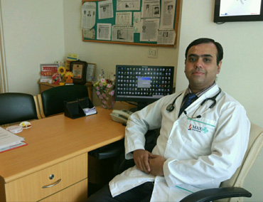
Heart and child clinic-F-87, Ashok vihar, Phase1, Delhi 110052
Timing: 7-9 am and 6-9 pm
Address :
Max superspeciality Hospital, Shalimar bagh
FC -50,Shalimar Bagh,Delhi 88 (C and D block,near Haiderpur metro station)Timing: 9 am to 5 pm
An Echocardiogram,
sometimes called an "echo", is an imaging test that uses the echoes of sound waves that are bounced off your heart to create a moving picture of your moving heart, its muscles and valves. It shows how well your heart is working, as well as how large your heart is. A small hand-held device called a transducer is used to transmit and receive the sound waves and echoes that take the pictures of your heart. Special equipment changes the echoes into images of your heart that are seen on a monitor. These images help the doctor evaluate your heart.
An echo is very safe and painless. In fact, echocardiograms are very similar to the imaging technique called sonograms or ultrasounds. Before your Echo - make sure you wear a two-piece outfit, you may need to undress from the waist up and put on a gown.
Although the test itself takes less than 45 minutes, you should allow extra time to check in. For this test, which is a non-stress echocardiogram, you may take your normal medications before the test. During your Echo Small electrodes are placed on your chest to monitor your heartbeat. A transducer coated with warm gel is moved firmly over your chest. This device creates the sound waves that make the images of your heart. At times, you may be asked to exhale and hold your breath for a few seconds. Air in your lungs can affect the images.
The images of your heart are recorded so your doctor can review them.
What is an Exercise Treadmill Test?
It is an electrocardiogram (EKG or ECG) conducted while walking on a treadmill to measure the heart's response to the stress of physical activity. Your blood pressure and heart rhythm are monitored during the test as well. The results aid in the detection of the disease which causes heart attacks, determination of cardiovascular fitness, and the safety of an exercise program. The test can show if there's a lack of blood supply through the arteries that go to your heart.
Before your exercise treadmill test Don't eat, drink, smoke, or have any caffeine for 3 hours before your test. Make sure you wear a two-piece outfit and walking shoes, you may need to undress from the waist up and put on a gown. U may need to discontinue some medications like beta blockers ,nitrates two days prior to test( only after consultation with your doctor).
What happens during the test? You are hooked up to equipment to monitor your heart. You walk slowly in place on the treadmill. The treadmill will tilt so you feel like you are going up a small hill. It changes speeds to make you walk faster in order to increase your heart rate. The test is over when you reach your maximum heart rate or you feel you need to complete the test.
After slowing down for a few minutes, you will sit down and your heart rate and blood pressure will be monitored. Report any symptoms Be sure to tell the doctor if you feel any of the following during the test: Chest, arm, or jaw discomfort Severe shortness of breath Fatigue Dizziness Leg cramps or soreness.
After your test As soon as the test is over, you may eat and return to your normal routine.
Carotid ultrasound is a safe, painless procedure that uses sound waves to examine the structure and function of the carotid arteries in the neck. You have two carotid arteries, one on each side of the neck, which deliver blood from the heart to the brain.
Carotid ultrasound can reveal whether an artery has any blockage and how well blood flows through the artery. Carotid ultrasound is usually used to screen for blockages that indicate an increased risk of stroke. Results from a carotid ultrasound can help your doctor determine what kind of treatment you may need to lower your risk.
The primary purpose of a carotid ultrasound is to screen for a narrowing of the carotid arteries that indicates an increased risk of stroke.
Narrowing is usually caused by plaques — a buildup of fats, cholesterol, calcium and other substances that circulate in the bloodstream. Early detection of narrowing of the carotid arteries enables your doctor to begin treatments that improve blood flow to the brain and decrease your risk of stroke.
Holter monitoring is used to record your heartbeat away from our office and allows your doctor to evaluate how it responds to normal activity. When you receive a Holter Test Monitor, small, painless electrodes (conducting patches) are placed on your chest and attached to a small recording monitor. The monitor can be placed in a pocket or small pouch worn around your neck. The monitor is battery operated. While you wear the monitor, it records your heart's electrical activity. You need to keep the device on at least 24 hours and complete a diary of your activities while wearing the monitor. After 24 hours, you return the monitor to our office. Your records will be studied to determine if there have been any irregular heart rhythms. It is very important that you accurately record your symptoms and activities so that the doctor can match them with your Holter monitor findings. There is no special preparation, except do not apply lotion to your chest before. Tell our staff if you are allergic to tape or other adhesives. This is a painless test. However, some people may need to have their chest shaved so the electrodes can stick. Tips while wearing the monitor: Do not get the monitor wet. This includes no shower or bath. Follow your normal routine, including exercise, work, etc. Keep a detailed diary of all activities during the monitoring time, making sure to note any symptoms you may feel and when you take your medications.
Angiography is a procedure in which a thin tube (catheter ) is inserted from wrist or groin and is threaded to arteries supplying heart. Next a special dye is injected and X-ray pictures are taken to visualize the blockage in vessels supplying the heart.The procedure is done under local anaesthesia, patient is awake and causes little or no pain..Coronary angiography rarely causes serious complications.
It is a procedure that is used to open or dilate an obstructed artery that leads to the heart. It is performed in same way and in combination with an angiogram via wrist/groin.The angioplasty is done by inserting a special tiny balloon through the catheter into the narrowed part of blood vessel.
The balloon is then slowly inflated. As the balloon inflates, it presses the plaque and in doing so opens the artery.At this point ,the stent is inserted. A stent is a soft mesh like piece of material that is placed into the narrowed part of artery. The stent acts as a scaffold or bridge to keep the artery open.
After the stent is inserted ,the balloon is removed.The stent remains in the artery to keep it open. There are two types of stents- drug coated and non drug coated. Nowadays mostly drug coated stents are implanted because reblockage rates are lesser with these.
Biodegradable stents are hailed as the next big thing in the treatment of blocked heart arteries .Just as a cast holds a broken bone in place while it heals, but is removed eventually, this new type of stent props an artery open and then dissolves in the body after two years. Future studies will be able to shed more light on safety and effectiveness of these absorbable stents compared to conventional stents.
If your heart is beating too slowly or you have been diagnosed with congestive heart failure, you may need to have a pacemaker inserted. A pacemaker is a small implanted device that regulates your heart beat electronically. It monitors your heart’s rhythm and generates a painless electric impulse that triggers a heart beat.
The procedure of implanting a pacemaker is done under local anaesthesia. A lead is placed into right heart chamber via veins of chest and is connected to a battery like device which is placed underneath skin of chest.
Special instructions after pacemaker implantation-
Hold cell phone upto ear that is opposite to site where pacemaker is implanted. Do not put your cell phone or MP3 player in shirt pocket over your pacemaker (if the devices are turned on)
You can still use household appliances (like microwave ovens) but avoid close and prolonged exposure
You can walk through security systems, metal detectors at normal pace. Notify security staff that you have a pacemaker, so that metal detector wand is not held for too long over your pacemaker site. Avoid sitting/Standing close to security systems of pacemaker
Medical procedures which can disrupt your pacemaker include – Magnetic Resonance Imaging(MRI), Shock wave Lithotripsy (for renal stones), electrocautery (to stop bleeding during surgery)
ICD is a special pacemaker that besides pacing a slow heart is capable of giving shock to treat life threatening ventricular arrhythmias, sudden cardiac death survivors and primary prevention of arrhythmias in heart attack survivors with weakened hearts.
Biventricular Pacemaker (CRT) is a pacemaker that is used to treat patients with heart failure. In some patients with advanced heart failure , delay in heart ventricle contractions occur.Heart failure means the heart’s pumping capacity is weaker than normal.The CRT pacing device (also called a biventricular pacemaker) is an electronically paced device that is surgically implanted under the skin.
The device has two or three leads (wires) that are positioned in the heart . CRT device helps the heart to beat in a more balanced way. CRT improves symptoms of heart failure, survival,quality of life,heart function, ability to exercise and decrease in hospitalizations in selected patients with heart failure.
Balloon mitral valvotomy is a procedure for opening of stenotic mitral valves due to rheumatic heart disease. This procedure is done under local anaesthesia.A balloon is advanced from groin to left side of heart and is inflated to open the valve. It is a good alternative to open heart surgery in patients whose valves are suitable for this procedure.
TAVR Usually valve replacement requires an open heart procedure with a "sternotomy", in which the chest is surgically separated (open) for the procedure. The TAVR or TAVI procedures can be done through very small openings that leave all the chest bones in place. This procedure is fairly new and is approved for people with symptomatic aortic stenosis who are considered a high risk patient for standard valve replacement surgery.
This minimally invasive surgical procedure repairs the valve without removing the old, damaged valve. Instead, it wedges a replacement valve into the aortic valve’s place. The surgery may be called a transcatheter aortic valve replacement (TAVR) or transcatheter aortic valve implantation (TAVI) Somewhat similar to a stent placed in an artery, the TAVI approach delivers a fully collapsible replacement valve to the valve site through a catheter which is inserted from groin.
Once the new valve is expanded, it pushes the old valve leaflets out of the way and the tissue in the replacement valve takes over the job of regulating blood flow.